Ramesa Shafi Bhat ![]() ,
Sooad Al-daihan
,
Sooad Al-daihan
For correspondence:- Ramesa Bhat Email: rbhat@ksu.edu.sa Tel:+9668055371
Received: 15 October 2015 Accepted: 12 March 2016 Published: 30 April 2016
Citation: Bhat RS, Al-daihan S. Liver injury from ampicillin-induced intestinal microbiota distresses in rats fed carbohydrate- and protein-rich diets. Trop J Pharm Res 2016; 15(4):709-716 doi: 10.4314/tjpr.v15i4.6
© 2016 The authors.
This is an Open Access article that uses a funding model which does not charge readers or their institutions for access and distributed under the terms of the Creative Commons Attribution License (http://creativecommons.org/licenses/by/4.0) and the Budapest Open Access Initiative (http://www.budapestopenaccessinitiative.org/read), which permit unrestricted use, distribution, and reproduction in any medium, provided the original work is properly credited..
Purpose: To investigate the effect of ampicillin on rat intestinal microflora and liver in the presence of high carbohydrate and protein diets.
Methods: Male Wistar albino rats were divided into four groups. The first group served as the control, the second group was treated with ampicillin (50 mg/kg for 3 weeks) and fed with a standard diet, while the third and fourth groups were treated with the same dose of ampicillin and fed with acarbohydrate- and protein-rich diets, respectively, to observe the effect of diet on gut flora and liver. Fecal specimens were collected and used for qualitative determination of gut microbiota composition. Serum hepato-specific markers (AST, ALT and ALP) were estimated. The antioxidant status of liver tissues was estimated for GSH, MDA, GST, LDH and vitamin C l, in addition to sodium and potassium.
Results: Administration of orogastric dose of ampicillin for 3 weeks induced inhibition of E.coli, yeasts, total anaerobes, and anaerobic lactobacilli with new growth of P. vulgaris and K. pneumonia. The levels of serum AST, ALT and ALP showed significant (p F6; 0.05) increase to 163, 112.38 and 115.35 %, respectively in ampicillin-treated animals, compared to control. Also significant (p F6; 0.05) increase in lipid peroxidation (120 %) and LDH (111 %) coupled with significant (p F6; 0.05) decrease in glutathione (74.57 %), vitamin C (63.49 %) and glutathione S-transferase (41.51 %) were observed in ampicillin-treated groups. No significant variation (p F6; 0.05) in sodium and potassium levels were found between control and the treated group after 3 weeks of treatment.
Conclusion: These results confirm that extended ampicillin therapy disrupts gut flora, which results in liver injury; hence, overuse of antibiotics should be avoid.
Introduction
Gut microbiota has significant effects on human health and disease, particularly nutrient uptake. However, the effects of gut microbiota are not limited to the intestine. In healthy individuals, all surfaces of the body exposed to the environment are normally inhabited by micro-organisms [1]. Microbiota performs vital functions essential to health maintenance, including food processing, digestion of complex indigestible polysaccharides and synthesis of vitamins [2]. Gut microbial flora has spatial and temporal complexity that varies from person to person, body niche, age, geographic location, health status, diet and type of host [3]. Even within the same individual, the composition of microbial flora can vary according to changes in diet, stress, sexual behavior, medication, hormonal changes, exposure to antibiotics and other host-related factors. When the mutually beneficial relationship between the flora and body is disrupted, some bacterial can overgrow in the intestine and cause disease [4].
The gut and liver are closely connected with each other through the products of digestion and absorption, bile, hormones and inflammatory mediators. The main blood supply to the liver is through direct venous outflow of the intestine from the portal vein, which makes the liver the first line of defense against gut-derived antigens, food antigens, toxins, microbial-derived products, and microorganisms [5]. Toxins produced by gut flora, such as ammonia, ethanol, acetaldehyde, phenols, and benzodiazepines are finally metabolized by the liver [6]. Thus, normal levels of gut flora are very essential for proper liver function and physiology.
Under normal conditions, the composition of the microbiota is relatively stable for long periods of time [6], but this can change due to antibiotic treatments. Overuse of antibiotics can cause ecological disruption in normal intestinal micro flora by allowing the resistant bacteria to overgrow with the diminution of beneficial bacteria [7]. Antibiotics are often used to treat a specific illness without considering how the antibiotic will affect the normal flora [7].
Diet is considered to be a major environmental factor affecting gut microbiota diversity and functionality [8]. Diet can also have a marked effect on the gut environment, including gut transit time and pH; and the composition of the microbiota [8]. The aim of this study was to characterize changes in intestinal microbiota induced by ampicillin and its effect on liver in the presence of a high carbohydrate and protein diet.
Methods
Animals and diet
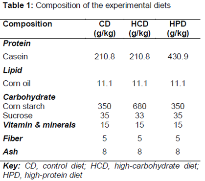
Adult male Wistar albino rats at the age of 5 weeks and 100 - 150 g body weight were purchased from the Animal House of Science College, King Saud University Riyadh. The animals were fed with a standard pellet diet, and water was provided ad libitum. The animals were maintained in a controlled environment under standard conditions of temperature and humidity with an alternating light and dark cycle. The protocols used in the present study was approved by the Ethics Committee at the King Saud University (approval ref no. 8/25/220358), and all experiments were performed according to the guidelines of National Animal Care and Use Committee [9]. The rats were randomly divided into four groups, in which a group consisted of ten rats. The first group served as a control. The second group received an orogastric dose of ampicillin (50 mg/kg for three weeks) [10]. Groups 3 and 4 were administered an orogastric dose of ampicillin (50 mg/kg for three weeks) [10] and converted to a high carbohydrate and high protein diet, respectively. shows details of diet composition. The diets were freshly prepared each day as pellets, and the ingredients were individually stored at 4 °C.
Sample preparation
Serum
Blood was collected from the retro-orbital plexus without the use of an anticoagulant. The blood was allowed to stand for 10 min before centrifugation at 2,000 rpm for 10 min to obtain serum for analysis of transaminases and alkaline phosphatase.
Tissue
Liver tissues isolated from the sacrificed animals were washed in ice-cold 1.15 % KCl and homogenized in a homogenizing buffer (50 mM Tris-HCl, 1.15 % KCl pH 7.4) using a Teflon homogenizer. The homogenate was centrifuged at 4000 × g for 20 min to obtain supernatant that was subsequently used for various biochemical assays.
Biochemical analyses
Serum alanine aminotransferase (ALT)
ALT was estimated by the method of Reitman and Frankel [11]. Briefly, 0.5 ml of substrate (2 mM α-ketoglutarate, 0.2 M DL-alanine in 0.1 M phosphate buffer pH 7.4) was incubated at 37 °C for 5 min. 0.1 ml of freshly prepared serum was added to the aliquot and again incubated at 37 °C for 30 min. At the end of the incubation, 0.5 ml of 2, 4-dinitrophenyl hydrazine was added, and the aliquot was incubated for 30 min at room temperature. Next, 0.5 ml of 0.4 N NaOH was added, and the aliquot was incubated for an additional 30 min. Absorbance was then recorded at 505 nm against water blank.
Serum aspartate aminotransferase (AST)
AST was estimated using the method of Reitman and Frankel [11]. However, the substrate was 2 mM α-ketoglutarate, 0.2 M DL-aspartate, and the rest of the procedure was similar to the ALT measurement method.
Serum alkaline phosphatase (ALP)
Serum alkaline phosphatase activity was measured according to the method of King and Armstrong [12] using disodium phenyl phosphate as a substrate. The developed color was read at 510 nm.
Measurement of lipid peroxidation
Lipid oxidation was evaluated by measuring the levels of lipid peroxidation by-products as thiobarbituric acid reactive substances (TBARS), namely malondialdehyde (MDA), using the method of Ruiz-Larrea et al [13].
Consistent with these findings, the samples were heated with TBA at a low pH, and the formation of a pink chromogen was measured at an absorbance of 532 nm. The concentration of lipid peroxides was calculated as μmoles/ml using the extinction coefficient of MDA.
Vitamin C assay
A vitamin C assay was performed according to the method of Jagota and Dani [14]. In this assay, 0.2 ml of liver homogenate was mixed with 0.8 ml of 10 % trichloroacetic acid (TCA) and incubated on ice for 5 min. The samples were then centrifuged for 10 min at 3500 rpm and 4 °C. Next, 1.5 ml double distilled water was subsequently added to 0.5 ml of the supernatant. Finally, 2 ml of Folin-phenol reagent was added, and the absorbance was measured at 760 nm after 10 min.
Assay of glutathione (GSH)
The GSH content was determined according to the method described by Beutler et al [15] using 5, 5′-dithiobis 2-nitrobenzoic acid (DTNB) with sulfhy-dryl compounds to produce a relatively stable yellow color.
Glutathione-S-transferase (GST) assay
The GST activity was determined using the method of Habig et al [16]. The 1-chloro-2-4-di-nitrobenzene (CDNB) was neutralized by an enzyme in the presence of GSH as a cosubstrate. The change in absorbance was measured at 340 nm, and the activity was expressed as nmol/min/mg protein.
Lactate dehydrogenase (LDH) assay
The quantitative determination of LDH in brain homogenates was performed using the lactate-to-pyruvate kinetic method described by Henry et al [17].
Determination of potassium levels
Potassium levels were measured in a protein-free alkaline medium by reaction with sodium tetraphenyl boron, which produced a colloidal suspension. The turbidity of such a suspension was proportional to the potassium concentrations in the range of 2–7 mmol/l [18].
Determination of sodium levels
Sodium levels were assayed by enzymatic determination of sodium, i.e., the measurement of sodium-dependent galactosidase activity using ortho-Nitrophenyl- β-galactoside (ONPG) as a substrate [19].
Microbiological examination
Cecal contents were collected in sterile tubes and immediately stored at -20 °C. The frozen samples were then analyzed. Fecal samples were cultured in aerobic, as well as anaerobic conditions, and were continuously monitored for one week. Positive cultures were plated on appropriate media, and species were identified using Scepter micro-dilution and standard bacteriological techniques. Identification of microorganisms were detected after 24 and 48 h of incubation at 37 oC.
Statistical analysis
The results are expressed as mean ± standard deviation (SD), and were evaluated by SPSS (version 12.0) and Origin 6 software using one-way ANOVA followed by Dunnett’s test for multiple comparisons. Differences were considered statistically significant at p < 0.05.
Results
Effect of ampicillin on intestinal microflora of rats fed with different diet
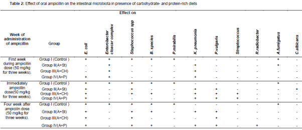
The qualitative changes of rat feces of the control and all treatment groups are summarized in Several variations were found in the fecal flora of rats in all treatment groups when compared with the control. Ampicillin induced inhibition of E. coli, yeasts, total anaerobes, and anaerobic lactobacilli after one week of treatment. The administration of ampicillin caused the disappearance of Enterobacter cloacar complex after one week and inhibited yeasts, total anaerobes, and anaerobic lactobacilli after three weeks of treatment. New colonies of P. vulgaris and K. pneumonia were found in all ampicillin-treated groups.
Effect of ampicillin on liver functions of rats fed with different diet
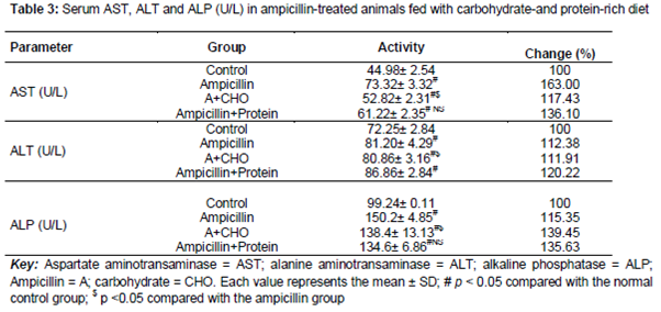
After treatment with ampicillin, the levels of serum AST, ALT and ALP significantly increased, compared with the control by approximately 163, 112.38 and 115.35 %, respectively (). Liver injury manifested by elevation of serum AST (117.43 % of control), ALT (111.91 % of control) and ALP (139.45 % of control) activities was observed in group III (ampicillin +carbohydrate-rich diet, ).
Rats treated with ampicillin in the presence of a protein-rich diet showed a significant increase in AST (136.10 % of control), ALT (120.22 % of control) and ALP (135.63 % of control) activities ().
Effect of ampicillin on oxidative stress of rats fed with different diet
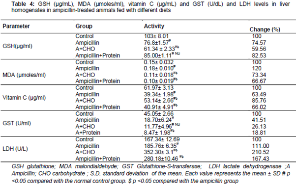
Antibiotic administration to rats showed a significant increase in MDA (120 % of control) and LDH (111 % of control) activities with a concomitant decrease of GST (41.51 % of control), GSH (74.57 % of control), and Vitamin C (63.49 % of control) in liver.
Compared to control groups, the antibiotic-treated rats with different diets also exhibited an increase in MDA and LDH activities with an associated decrease of GST, GSH, and Vitamin C ().
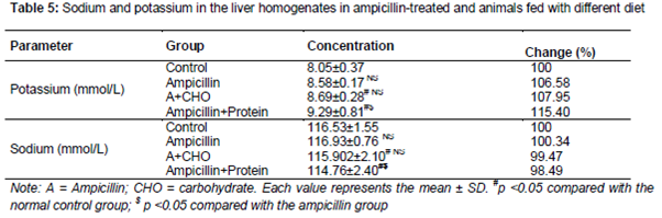
Antibiotic-induced hepatotoxicity associated with changes in liver sodium and potassium content
The changes in sodium and potassium content in all treated groups are presented in . Administration of antibiotic resulted in an increase in sodium content of the rat liver by 100.34, and a decrease in the presence of a carbohydrate and protein diet with 99.47 and 98.49 %, respectively. Potassium levels were increased in all antibiotic-treated groups by 106.58, 107.95 and 115.40 % compared with the control group ().
Discussion
Oral administration of antibiotics usually disrupts the ecological balance of gut flora in body. Oral administration of ampicillin among the all of the groups in the present study resulted in an imbalance in the composition of gut microbiota with the decrease in the number of E. coli, yeasts, total anaerobes, and anaerobic lactobacilli and increase in the number of Klebsiella and Proteus species. These results were consistent with the previous work of Knothe & Wiedemann [20] in which they reported the overgrowth of ampicillin-resistant Citrobacter, Klebsiella and Proteus species with a decrease in E. coli, enterococci, bifidobacteria and anaerobic Gram-negative rods after five days treatment with ampicillin in humans. Interestingly, a recent study also highlighted the overgrowth of K. pneumonia with ampicillin treatment in mice [21]. K. pneumonia strains have the ability to cross the intestinal barrier and cause liver abscess [21].
Antibiotics are considered as a common cause of drug-induced liver injury with only mild hepatic impairment [7]. In the present investigation, we observed that ampicillin induced liver toxicity in rats. This was evident from the liver function test as the serum concentration of AST, ALT and ALP significantly increased in all of the treated groups.
Oxidative stress and lipid peroxidation that are mediated by oxygen-free radicals have been implicated as a common link between chronic liver damage and hepatic fibrosis [22]. The results demonstrated that administration of ampicillin resulted in a markedly significant increase in MDA and LDH activities with a concomitant decrease in GST, GSH and Vitamin C in liver. These results were consistent with some previous studies [23] demonstrating that carbohydrate- and protein-rich diets were unable to normalize the elevated hepatic markers or elevated biochemical oxidative stress markers in liver (Tables 3 and 4). Diet is considered to be a major environmental factor influencing gut microbiota diversity and functionality [8].
Similarly, products of the intestinal microbiota have a direct effect on hepatic function [8]. The majority of gut flora metabolites are hepatotoxic and are metabolized in the liver only [8]. Dietary protein is metabolized into harmful nitrogenous metabolites by gut flora via fermentation [24]. Carbohydrates activate a wide range of microbial enzymes and increase glucose flux to hepatic cells, which in turn produce short chain fatty acid in the liver, resulting in fatty liver diseases [25]. Alterations in gut bacteria also affect intestinal permeability and inflammation, thus also affecting metabolic disorders [24].
The active transport of sodium-potassium across the cell membrane is controlled by sodium-potassium-adenosine triphosphatase (Na+-K+-ATPase), which is an integral plasma membrane protein responsible for a large part of energy consumption, constituting the cellular metabolic rate. Na+-K+-ATPase controls cell volume, nerve and muscle signals and drives the transport of amino acids and sugars [26]. However, there was no significant difference from the control value with the administration of ampicillin in all treated groups, which may be an indication that it may be of low toxicity to the monovalent ion.
Conclusion
Antibiotic treatment, combined with carbohydrate and protein diets, increases liver toxicity. Their overuse leads to overabundance of modern day health challenges. Dietary changes can dramatically alter the balance of bacteria in the gut, which in turn can affect liver function.
Declarations
Acknowledgement
References
Archives


News Updates